Indexed in
Archive
-

Vol. 69 No. 4 (2024)
Cover Image
Two colorimetric methods, enzymatic (red) and non-enzymatic (yellow), for measuring the concentration of lactic acids in complex biological mixtures were compared.
Photo by Kseniya Vereshchagina (see page 203). -
Vol. 69 No. 3 (2024)
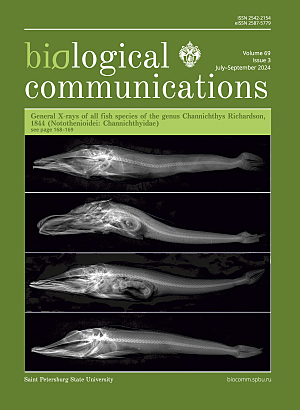
Cover Image
The most important diagnostic features of fish species of the genus Channichthys, seen on X-rays, such as: the number of rays in the first (D1) and second (D2) dorsal fins, pectoral fins (P), anal fin (A), the total number of vertebrae in the spine (vert.).
Photo by Ekaterina Nikolaeva (see page 162). -
Vol. 69 No. 2 (2024)
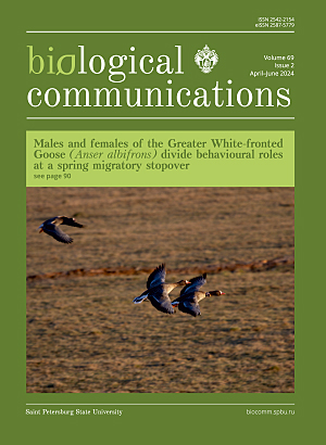
Cover Image
A pair of Greater White-fronted geese in the Kologriv Floodplain protected area, the female is marked with a black neckband SH5, 2022.
Photo by Petr Glazov (see page 90). -
Vol. 69 No. 1 (2024)
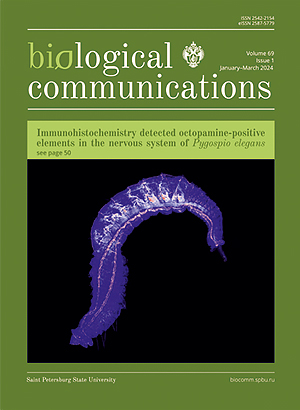
Cover Image
Confocal micrograph of the nervous system of regenerating annelid Pygospio elegans Claparede, 1863.
Photo by Zinaida Starunova (see page 50). -
Vol. 68 No. 4 (2023)
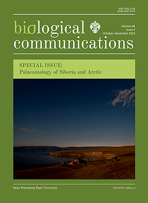
Cover Image
The camp of a paleontological expedition excavating the Miocene Tagay locality known for the findings of various vertebrates, including birds and mammals. Tagay Bay, the Olkhon Island of the Lake Baikal, Siberia, Russia.
Photo by Alexander Sizov (see page 273). -
Vol. 68 No. 3 (2023)
Cover Image
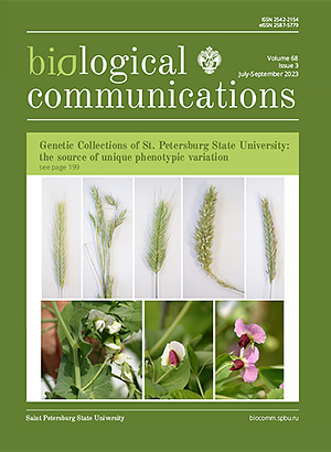
Variability of ear shape in rye (Secale cereale) lines of the Peterhof genetic collection of rye (top row) and garden pea (Pisum sativum) corolla color (bottom row) of the educational collection of the Department of Genetics and Biotechnology (St. Petersburg State University).
Photo by Pavel Zykin (see page 199). -
Vol. 68 No. 2 (2023)
Cover Image
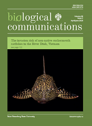
The suckermouth catfish Pterygoplichthys spp., rapidly and successfully invading tropical freshwaters around the world. The River Dinh, Khánh Hòa Province, Vietnam.
Photo by Dmitry Zworykin (see page 122). -
Vol. 68 No. 1 (2023)
Cover Image
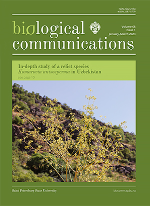
Komarovia anisosperma, a rare and endemic species of the Zarafshan Ridge, in its natural habitat. Kitab Geological State Reserve, Kashkadarya Region, Uzbekistan.
Photo by Ulugbek Kodirov (see page 10). -
Vol. 67 No. 4 (2022)
Cover Image
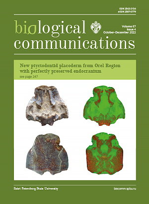
PIN 3725/1224, holotype of ptyctodontid Neruchella eichwaldi gen. et sp. nov. from the Famennian of Orel Region, Russia. Photograph and microtomographic images of the head shield.
Photo by Alexander Ivanov (see page 247). -
Vol. 67 No. 3 (2022)
Cover Image
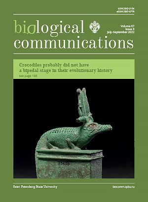
Although the ancient Egyptian god Sobek is usually depicted as an anthropomorphic bipedal creature with a crocodile head, there are quadrupedal sculptures of him.
Bronze figure of ancient Egyptian deity Sobek, EA37450, The British Museum, https://www.britishmuseum.org/collection/object/Y_EA37450 (see page 168). -
Vol. 67 No. 2 (2022)
Cover Image
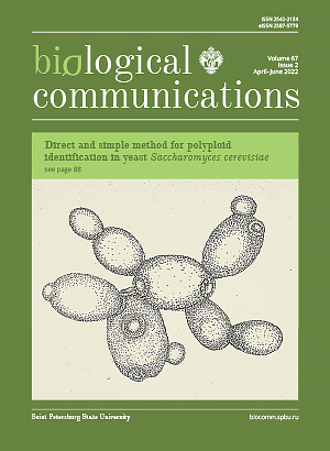
Proliferating cells of yeast Saccharomyces cerevisiae: maternal cells with scars and young buds.
Drawing by Yulia Andreychuk (see page 88). -
Vol. 67 No. 1 (2022)
Cover Image
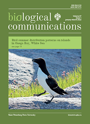
Razorbill (Alca torda) — a colonial species that breeds mainly on small islands. Onega Bay, White Sea, Russia.
Photo by Maria Matantseva (see page 19). -
Vol. 66 No. 4 (2021)
Cover Image
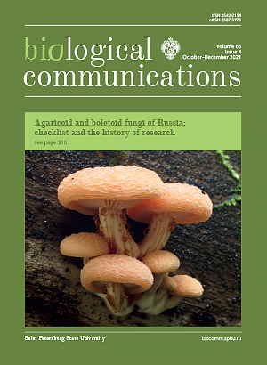
Basidiomata of Rhodotus palmatus, a species assessed for the IUCN Red List. Suma River valley, Leningrad Oblast, Russia.
Photo by Lyudmila Kalinina (see page 316). -
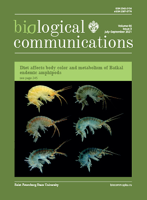
Vol. 66 No. 3 (2021)
Cover Image
Natural variety of coloration of the endemic Baikal amphipod species Eulimnogammarus cyaneus (Dybowsky, 1874).
Photo by Polina Drozdova (see page 245). -
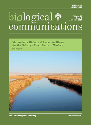
Vol. 66 No. 2 (2021)
Cover Image
The valley of the Sakarya River near Balıkdamı Wildlife Development Area. Eskişehir Province, Turkey.
Photo by Arda Acemi (see page 151). -
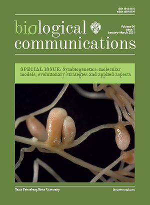
Vol. 66 No. 1 (2021)
Cover Image
Nodules on the roots of the legume plant Hedysarum arcticum growing on Kurungnakh Island. Ust-Lensk State Nature Reserve, Lena Delta, Yakutia, North-Eastern Siberia, Russia.
Photo by Denis Karlov. -
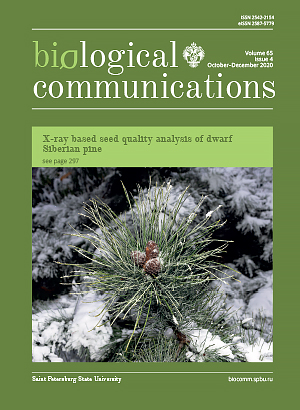
Vol. 65 No. 4 (2020)
Cover Image
Dwarf Siberian pine Pinus pumila at Botanical Garden of Peter the Great. Komarov Botanical Institute, Russian Academy of Sciences, Saint Petersburg, Russia.
Photo by Roman Khalikov (see page 297). -
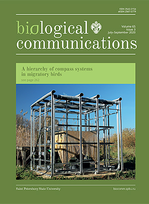
Vol. 65 No. 3 (2020)
Cover Image
The master of magnetic field. The double-wrapped, three-dimensional Merritt four-coil magnetic system which is used to study a hierarchy of compass systems in migratory birds. Biostation Rybachy, Kaliningrad Region, Russia.
Photo by Alexander Pakhomov (see page 262). -
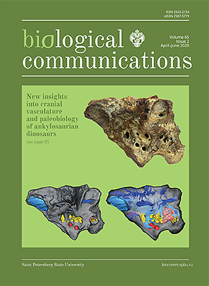
Vol. 65 No. 2 (2020)
Cover Image
ZIN PH 1/16, holotype of Bissektipelta archibaldi (Dinosauria: Ankylosauridae) from the Bissekty Formation (Turonian), Uzbekistan. Photograph and semitransparent CT-based model showing the braincase and its endocast contents.
Photo by Ivan Kuzmin (see page 85). -
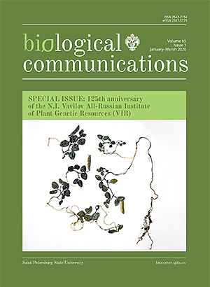
Vol. 65 No. 1 (2020)
Cover Image
Herbarium specimen of Vavilovia formosa from the Ragdan-chai Gorge, Dagestan, 2660–2700 m a.s.l., collected on August 31, 1973 (VIR).
Photo by Margarita Vishnyakova (see page 28). -
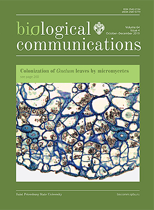
Vol. 64 No. 4 (2019)
Cover Image
Micrograph of transverse sections of developing cork warts in Gnetum gnemon.
Photo by Ianina Bogdanova (Pagoda) (see page 260). -
Vol. 64 No. 3 (2019)
Cover Image
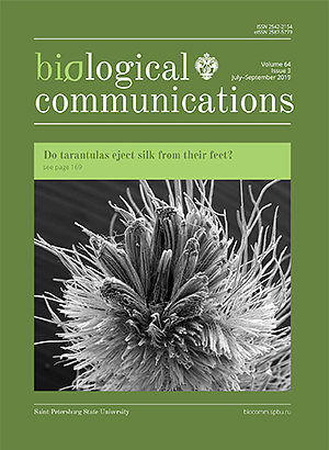
Scanning electron micrographs of distal tips of claw tufts setae of tarantulas.
Photo by Jaromír Hajer (see page 169). -
Vol. 64 No. 2 (2019)
Cover Image
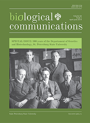
Dr. Yury Philipchenko (1882–1930), Professor, the first Head of Department of Genetics and Biotechnology (former Genetics and Experimental Zoology) of St. Petersburg State University and his colleagues (from left to right: seated — Th. G. Dobzhansky, Yu. A. Filipchenko, Ya. Ya. Lus; standing — V. I. Saveliev, N. N. Medvedev).
Photo (1926) from the archive of Department of Genetics and Biotechnology of St. Petersburg State University. -
Vol. 64 No. 1 (2019)
Cover Image
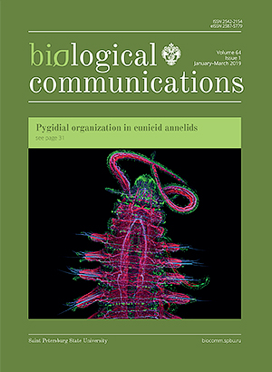
Confocal micrograph of the nervous system and musculature of the annelid Schistomeringos japonica (Annenkova, 1937), https://www.zin.ru/projects/neuromorphology/single_series_en.html?id=8.
Photo by Viktor Starunov (see page 31). -
Vol. 63 No. 4 (2018)
Cover Image
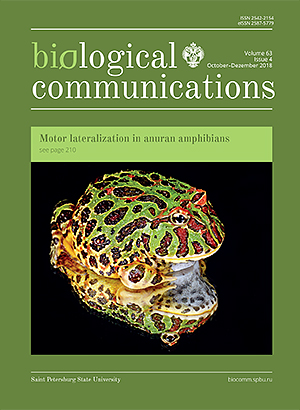
Close-up macro of an ornate horned frog (Ceratophrys ornata) reflected. Photo credit: Karen Arnold, https://www.publicdomainpictures.net/en/view-image.php?image=38773 (see page 210).


