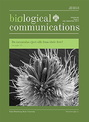
Claw tuft setae of tarantulas (Araneae, Mygalomorphae, Theraphosidae) and the production of fibrous materials. Do tarantulas eject silk from their feet?

Authors

  • Jaromír Hajer Department of Biology, J.E. Purkinje University, České mládeže 8, 400 96 Ústí nad Labem, Czech Republic https://orcid.org/0000-0002-2237-5175
  • Dana Řeháková Department of Biology, J.E. Purkinje University, České mládeže 8, 400 96 Ústí nad Labem, Czech Republic https://orcid.org/0000-0002-4228-967X

DOI:

https://doi.org/10.21638/spbu03.2019.301

Abstract

The study focused on the specialised tarsal setae of tarantulas Avicularia metallica and Heteroscodra maculata, and the fibrous and non-fibrous material produced by them. When irritated spiders moved along smooth, perpendicularly-oriented glass walls not covered in silk, the claw tuft setae, located at the tips of the tarsal segments, left behind footprints containing two types of fibrous material. Using electron scanning microscopy, it was discovered that these represent fragments of parallelly oriented bundles of hollow fibres forming the shafts of the setae and their lateral branches (1), as well as clusters of contracted nanofibrils which aggregated at the ends of these fibres (2). During climbing, this fibrous material was detected both on the substratum on which the spiders were moving, and also on their claw tuft setae. The climbing activity of irritated tarantulas is also associated with the secretion of a fluid which dries on contact with air. This secretion acts as an adhesive and facilitates the movement of tarantulas on smooth surfaces, but while doing so it also glues together the distal, lamellar parts of the groups of setae which are in contact with the substratum during climbing. There are no such claw tuft setae morphological changes observed in undisturbed tarantulas, moving freely around their tube-like shelters and on the surfaces of objects covered with silk which they have produced. The sources of the air-drying secretion are probably the tubular fibres forming the shafts of pretarsal setae. The bundles of hollow fibres are an example of a system that produces secretions via a surficial pathway. The spinnerets and silk-producing glands associated with them, located in the opisthosoma, represent a system that produces silk via a systemic pathway. However, the results of observational studies have not confirmed the ability of tarantulas’ feet to produce silk fibres of the same, or at least similar ultrastructure to that of the silk fibres produced by the activity of spinnerets and spinneret-associated silk glands.

Keywords:

spiders, adhesive setae, claw tufts, tarsal secretions, tarantula silk

Downloads

Download data is not yet available.
 

References

Arzt, E., Gorb, S., and Spolenak, R. 2003. From micro to nano contacts in biological attachment devices. Proceedings of the National Academy of Sciences 100(19):10603–10606. https://doi.org/10.1073/pnas.1534701100

Bond, J. E. 1994. Seta-spigot homology and silk production in 1st instar Antrodiaetus-unicolor spiderlings (Araneae, Antrodiaetidae). Journal of Arachnology 22(1):19–22.

Craig, C. L. 1997. Evolution of arthropod silks. Annual Review of Entomology 42:231–267. https://doi.org/10.1146/annurev.ento.42.1.231

Charpentier, P. 1992. Heterscodra maculata. British Tarantula Society Journal 8(2):11–19.

Dawydoff, C. 1949. Développement embryonnaire des arachnides.; 320–369 in: Traité de Zoologie, Anatomie, Systematique, Biologie, edited by Grassé, P. P. Paris: Masson.

Dunlop, J. A. 1995. Movements of scopulate claw tufts at the tarsus tip of a tarantula spider. Netherlands Journal of Zoology 45(3–4):513–520. https://doi.org/10.1163/156854295X00447

Eberhard, W. G. 2010. Possible functional significance of spigot placement on the spinnerets of spiders. Journal of Arachnology 38(3):407–414. https://doi.org/10.1636/b09-97.1

Federle, W. 2006. Why are so many adhesive pads hairy? Journal of Experimental Biology 209(14):2611–2621. https://doi.org/10.1242/jeb.02323

Ferretti, N., Pompozzi, G., Copperi, S., Wehitt, A., Galindez, E., Gonzalez, A., and Perez-Miles, F. 2017. A comparative morphological study of the epiandrous apparatus in mygalomorph spiders (Araneae, Mygalomorphae). Micron 93:9–19. https://doi.org/10.1016/j.micron.2016.11.005

Foelix, R. 2011. Biology of Spiders. 3 ed. New York: Oxford University Press.

Foelix, R., Erb, B., and Michalik, P. 2010. Scopulate hairs in male Liphistius spiders: probable contact chemoreceptors. Journal of Arachnology 38(3):599–603. https://doi.org/10.1636/b10-42.1

Foelix, R., Erb, B., and Rast, B. 2013. Alleged silk spigots on tarantula feet: Electron microscopy reveals sensory innervation, no silk. Arthropod Structure & Development 42(3):209–217. https://doi.org/10.1016/j.asd.2013.02.005

Foelix, R. F. and Chu-Wang, I. W. 1975. The structure of scopula hairs in spiders. Paper read at the 6th International Congress of Arachnology at Amsterdam.

Foelix, R. F., Rast, B., and Peattie, A. M. 2012. Silk secretion from tarantula feet revisited: alleged spigots are probably chemoreceptors. Journal of Experimental Biology 215(7):1084–1089. https://doi.org/10.1242/jeb.066811

Garb, J. 2013. Spider silk. An ancient biomaterial for 21st century research.; 252–281 in: Spider Research in the 21st Century: trends &, perspectives, edited by Penney, D.: Siri Scientific Press.

Glatz, L. 1972. Der Spinnapparat haplogyner Spinnen (Arachnida, Araneae). Zeitschrift für Morphologie der Tiere 72:1–26. https://doi.org/10.1007/BF00281752

Glatz, L. 1973. Der Spinnapparat der Orthognatha (Arachnidae, Araneae). Zeitschrift für Morphologie der Tiere 75:1–50. https://doi.org/10.1007/BF00723667

Gorb, S. N., Niederegger, S., Hayashi, C. Y., Summers, A. P., Voetsch, W., and Walther, P. 2006. Silk-like secretion from tarantula feet. Nature 443(7110):407–407. https://doi.org/10.1038/443407a

Hajer, J., Karschová, S., and Řeháková, D. 2016. Silks and silk-producing organs of Neotropical tarantula Avicularia metallica (Araneae, Mygalomorphae, Theraphosidae). Ecologica Montenegrina 7:313–327.

Haupt, J. 2003. The Mesothelaea — monograph of an exception group of spiders (Araneae: Mesothelae). Morphology, behaviour, ecology, taxonomy, distribution and phylogeny. Zoologica 154:1–102.

Haupt, J. and Kovoor, J. 1993. Silk-gland system and silk production in Mesothelae (Araneae). Annales Des Sciences Naturelles-Zoologie Et Biologie Animale 14(2):35–48.

Hilbrant, M. and Damen, W. G. M. 2015. The embryonic origin of the ampullate silk glands of the spider Cupiennius salei. Arthropod Structure & Development 44(3):280–288. https://doi.org/10.1016/j.asd.2015.04.001

Jocqué, R. and Dippenaar-Schoeman, A. S. 2006. Spider Families of the World. Koninklijk Museum voor Midden-Afrika.

Kautzch, G. 1910. Über die Entwicklung von Agelena labyrinthica Clerk. Zoologische Jahrbücher. Abteilung für Anatomie und Ontogenie der Tiere 28:30–36.

Kovoor, J. 1987. Comparative structure and histochemistry of silk-producing organs in Arachnids; 160–186 in: Ecophysiology of Spiders, edited by Nentwig, W. Berlin, Heidelberg: Springer Berlin Heidelberg. https://doi.org/10.1007/978-3-642-71552-5_12

Kropf, C. 2013. Hydraulic system of locomotion; 43–56 in: Spider Ecophysiology, edited by Nentwig, W. Berlin, Heidelberg: Springer Berlin Heidelberg. https://doi.org/10.1007/978-3-642-33989-9_4

Labarque, F. M., Wolff, J. O., Michalik, P., Griswold, C. E., and Ramirez, M. J. 2017. The evolution and function of spider feet (Araneae: Arachnida): multiple acquisitions of distal articulations. Zoological Journal of the Linnean Society 181(2):308–341. https://doi.org/10.1093/zoolinnean/zlw030

Lopez, A. and Emerit, M. 1988. New data on the epigastric apparatus of male of spiders. Bulletin of the British Arachnological Society 7(7):220–224.

Marples, B. J. 1967. The spinnerets and epiandrous glands of spiders. Zoological Journal of the Linnean Society 46(310):209–222. https://doi.org/10.1111/j.1096-3642.1967.tb00504.x

Miller, G. L., P. R., M., and A. R., B. 1988. Adhesive hairs in lycosid spiders of various life styles, including the occurence of claw tufts in Lycosa hentzi Banks. Bulletin of the British Arachnological Society 7(7):213–216.

Neretin, N. Y. 2016. The Morphology and ultrastructure of “Amphipod silk” glands in Ampithoe rubricata (Crustacea, Amphipoda, Ampithoidae). Biology Bulletin 43(7):628–642. https://doi.org/10.1134/s106235901607013x

Niederegger, S. 2013. Functional aspects of spider scopulae; 57–66 in: Spider Ecophysiology, edited by Nentwig, W. Berlin, Heidelberg: Springer Berlin Heidelberg. https://doi.org/10.1007/978-3-642-33989-9_5

Niewiarowski, P. H., Stark, A. Y., and Dhinojwala, A. 2016. Sticking to the story: outstanding challenges in gecko-inspired adhesives. Journal of Experimental Biology 219(7):912–919. https://doi.org/10.1242/jeb.080085

Osaki, S. 1996. Spider silk as mechanical lifeline. Nature 384(6608):419–419. https://doi.org/10.1038/384419a0

Peattie, A. M., Dirks, J. H., Henriques, S., and Federle, W. 2011. Arachnids secrete a fluid over their adhesive pads. Plos One 6(5). https://doi.org/10.1371/journal.pone.0020485

Pechmann, M., Khadjeh, S., Sprenger, F., and Prpic, N. M. 2010. Patterning mechanisms and morphological diversity of spider appendages and their importance for spider evolution. Arthropod Structure & Development 39(6):453–467. https://doi.org/10.1016/j.asd.2010.07.007

Perez-Miles, F., Perafan, C., and Santamaria, L. 2015. Tarantulas (Araneae: Theraphosidae) use different adhesive pads complementarily during climbing on smooth surfaces: experimental approach in eight arboreal and burrower species. Biology Open 4(12):1643–1648. https://doi.org/10.1242/bio.013144

Perez-Miles, F., Panzera, A., Ortiz-Villatoro, D., and Perdomo, C. 2009. Silk production from tarantula feet questioned. Nature 461(7267):E9. https://doi.org/10.1038/nature08404

Rind, F. C., Birkett, C. L., Duncan, B.-J. A., and Ranken, A. J. 2011. Tarantulas cling to smooth vertical surfaces by secreting silk from their feet. Journal of Experimental Biology 214(11):1874–1879. https://doi.org/10.1242/jeb.055657

Rovner, J. S. 1978. Adhesive hairs in spiders: behavioral functions and hydraulically mediated movement. Symposia of the Zoological Society of London 42:99–108.

Seyfarth, E. A. and Pfluger, H. J. 1984. Proprioreceptor distribution and control of muscle-reflex in the tibia of spider legs. Journal of Neurobiology 15(5):365–374. https://doi.org/10.1002/neu.480150506

Shultz, J. W. 1987. The origin of the spinning apparatus in spiders. Biological Reviews of the Cambridge Philosophical Society 62(2):89–113. https://doi.org/10.1111/j.1469-185X.1987.tb01263.x

Sutherland, T. D., Young, J. H., Weisman, S., Hayashi, C. Y., and Merritt, D. J. 2010. Insect silk: one name, many materials. Annual Review of Entomology 55(1):171–188. https://doi.org/10.1146/annurev-ento-112408-085401

Tirrell, D. A. 1996. Putting a new spin on spider silk. Science 271(5245):39–40. https://doi.org/10.1126/science.271.5245.39

Whitehead, W. F. and Rempel, J. G. 1959. A study of the musculature of the black window spider, Latrodectus mactans (Fabr.). Canadian Journal of Zoology 37(6):831–870. https://doi.org/10.1139/z59-084

Wolff, J. O. and Gorb, S. N. 2012. Comparative morphology of pretarsal scopulae in eleven spider families. Arthropod Structure & Development 41(5):419–433. https://doi.org/10.1016/j.asd.2012.04.004

Wolff, J. O. and Gorb, S. N. 2015. Adhesive foot pads: an adaptation to climbing? An ecological survey in hunting spiders. Zoology 118(1):1–7. https://doi.org/10.1016/j.zool.2014.04.006

Wolff, J. O., Nentwig, W., and Gorb, S. N. 2013. The great silk alternative: multiple co-evolution of web loss and sticky hairs in spiders. Plos One 8(5). https://doi.org/10.1371/journal.pone.0062682

World Spider Catalog. Version 20.0 2019. Natural History Museum Bern 2019 [cited 15. 6. 2019]. Available from http://wsc.nmbe.ch

Yoshicura, M. 1955. Embryological studies on the liphistiid spider Heptathel kimurai. Kumamoto Journal of Science Series B 2/3:41–48.

Downloads

Published

2019-10-30

How to Cite

Hajer, J., & Řeháková, D. (2019). Claw tuft setae of tarantulas (Araneae, Mygalomorphae, Theraphosidae) and the production of fibrous materials. Do tarantulas eject silk from their feet?. Biological Communications, 64(3), 169–182. https://doi.org/10.21638/spbu03.2019.301

Issue

Section

Full communications